Growers of the
"Périgord Black Truffle"


|
Growers of the "Périgord Black Truffle"
|
|
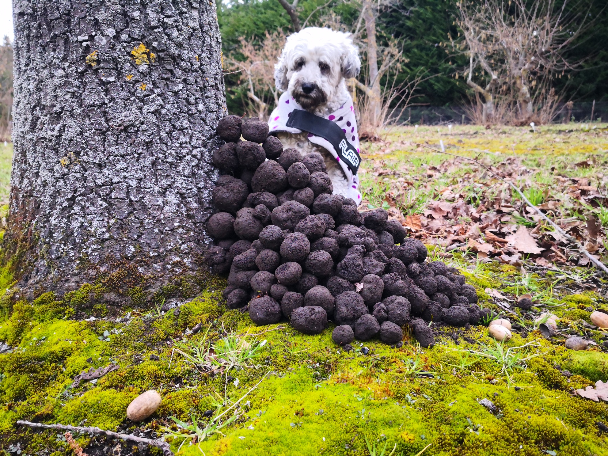
Report May 2014 Detection of Tuber melanosporum ectomycorrhizae on root samples from a Black Truffle plantation at 202 Old West Coast RoadOur client, Wayne Tewnion, was interested to test his own 18-year-old truffle plantation on the Old West Coast Road (Christchurch) to see if the presence of the target truffle species (Tuber melanosporum) could be detected or not. Wayne randomly selected 10 trees distributed over his 580-tree property and recorded the coordinates of the sampled trees. Wayne provided the following root samples to be tested by morphological analyses at the Plant & Food Research (PFR) Lincoln Mycorrhiza Laboratory: Oak #1, Oak #2, Oak #3, Oak #4, Oak #5 Conclusion * Only two root pieces from this sample were looked at. The piece 1 itself
held more than c. 90% of the ectomycorrhizae present in the whole sample. With
the naked eye, the few remaining pieces looked exactly the same as the two
pieces analysed.
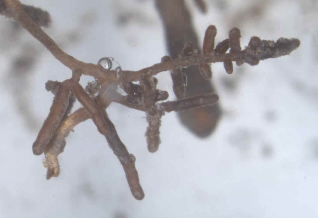
Figure 1. Dissecting microscope views (x10 magnification left, x7 right) of ectomycorrhizae characteristic of Tuber melanosporum on root pieces of sample from Oak #1.
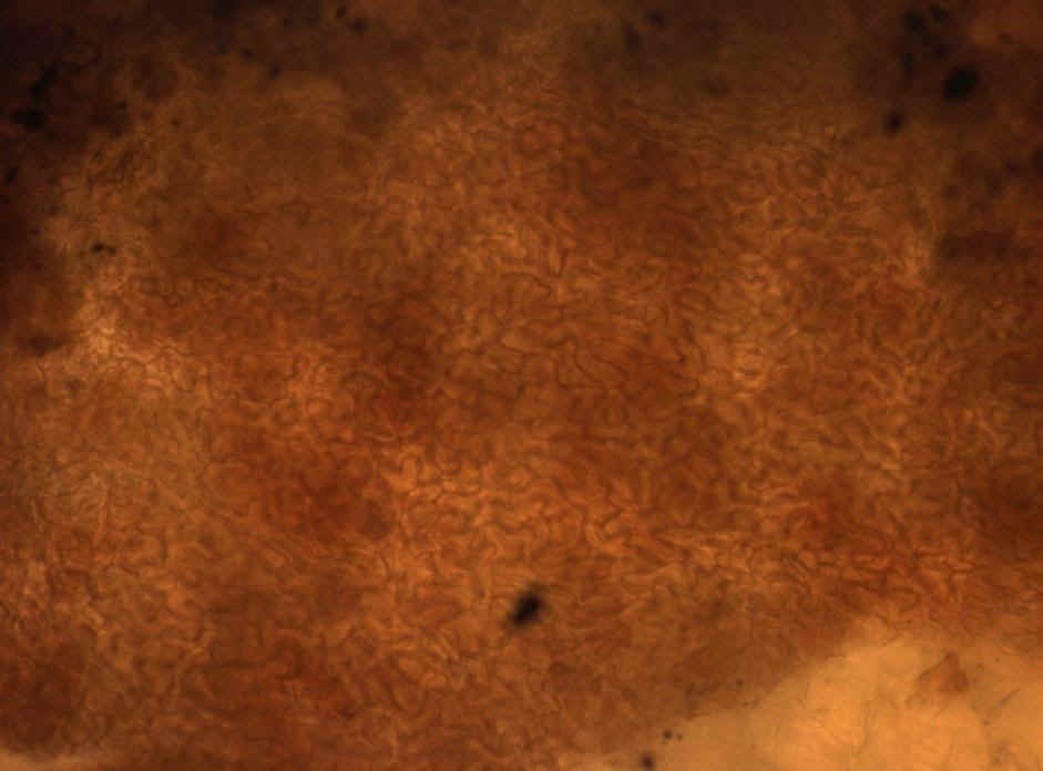
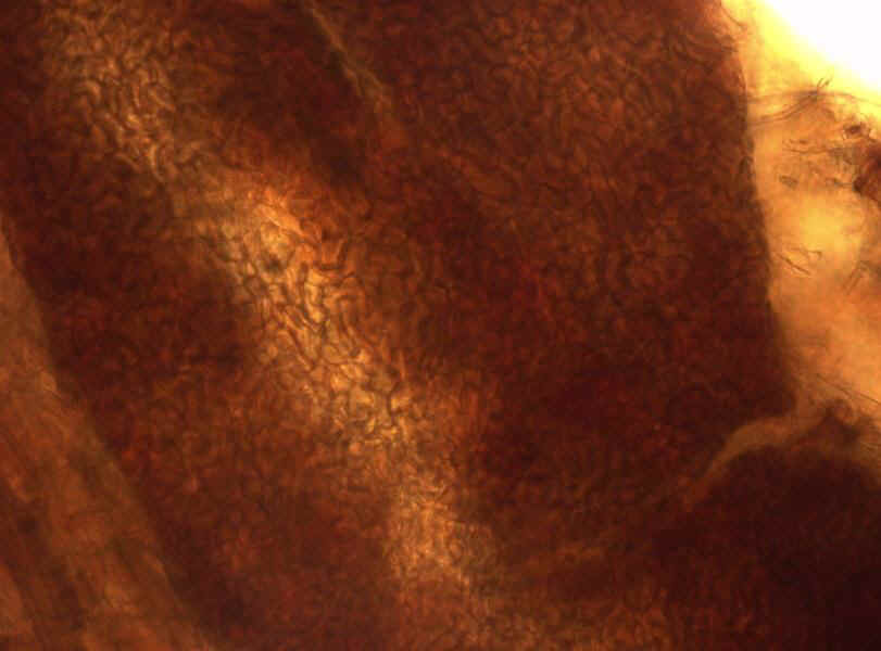
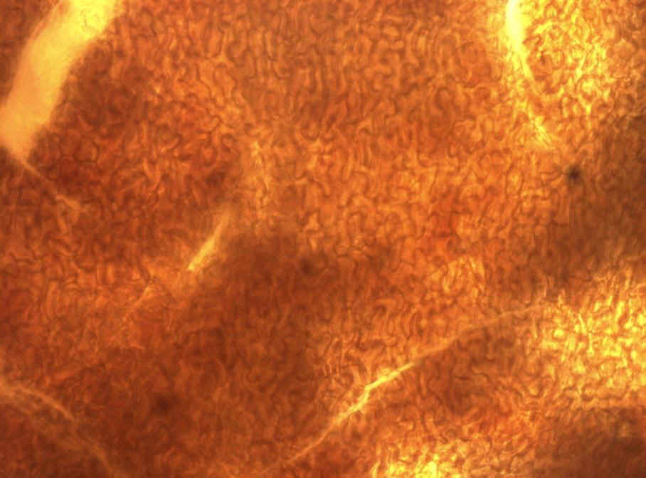
Figure 2. Compound microscope view (x400) of the ‘jigsaw puzzle’ pattern of the mantle of an ectomycorrhiza of T. melanosporum collected from Oak #1. This pattern is characteristic of several Tuber species, including T. melanosporum.
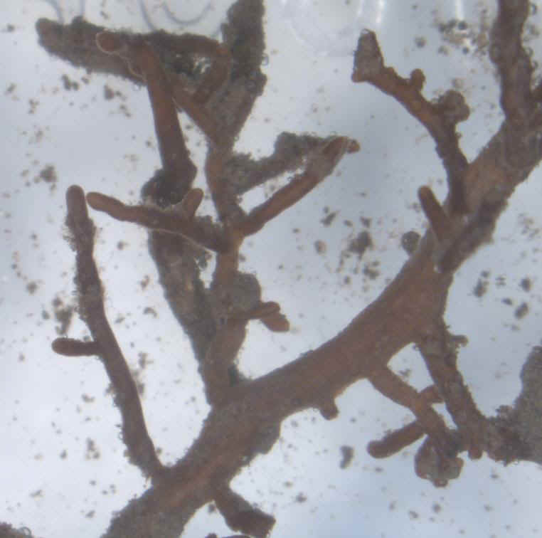
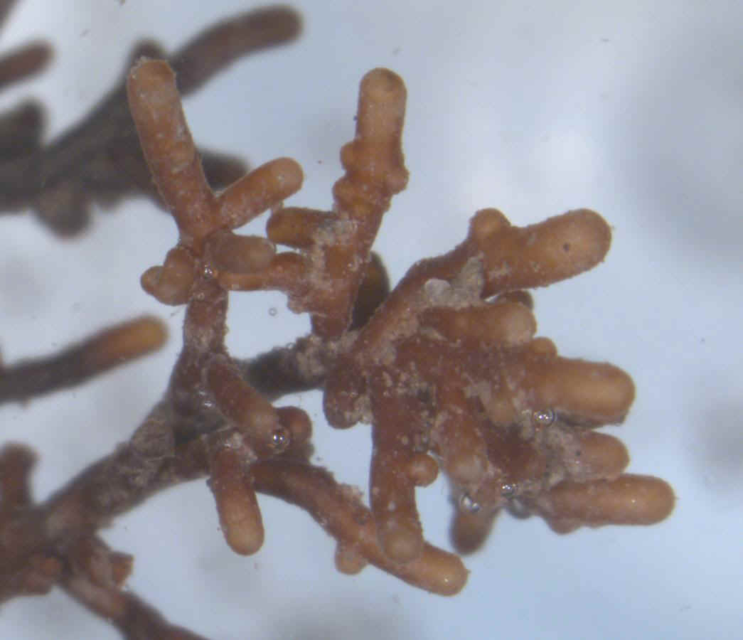
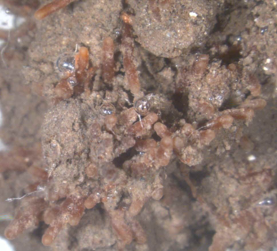
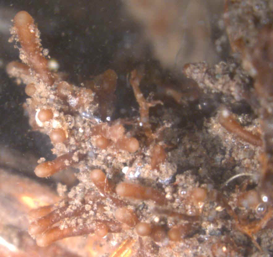
Figure 3. Dissecting microscope views of ectomycorrhizae characteristic of Tuber melanosporum on Oak #2 (x10 top left, x7 top right, x12 bottom) . Note the presence of clusters of ectomycorrhizae (Top).
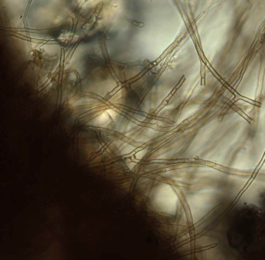
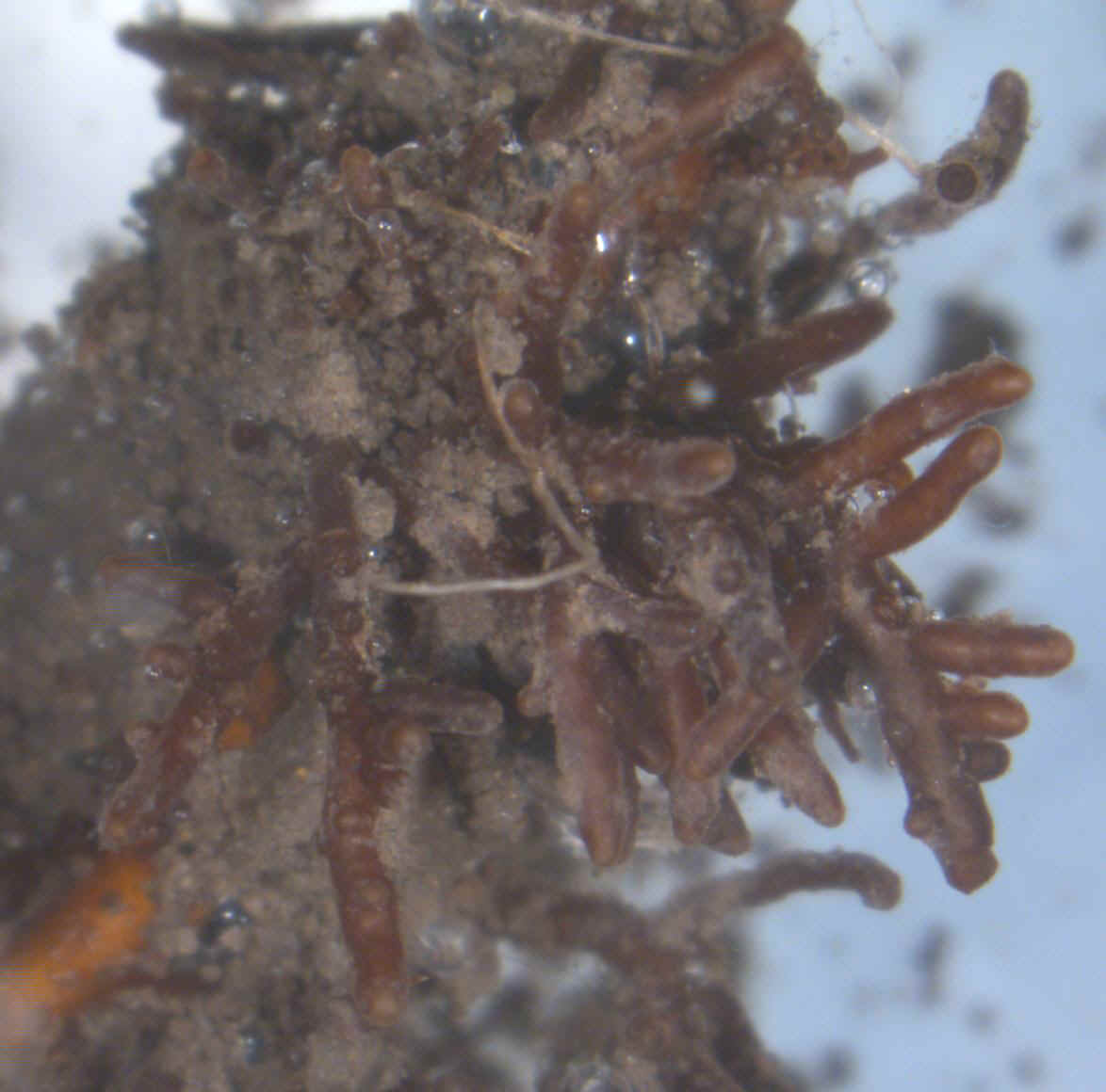
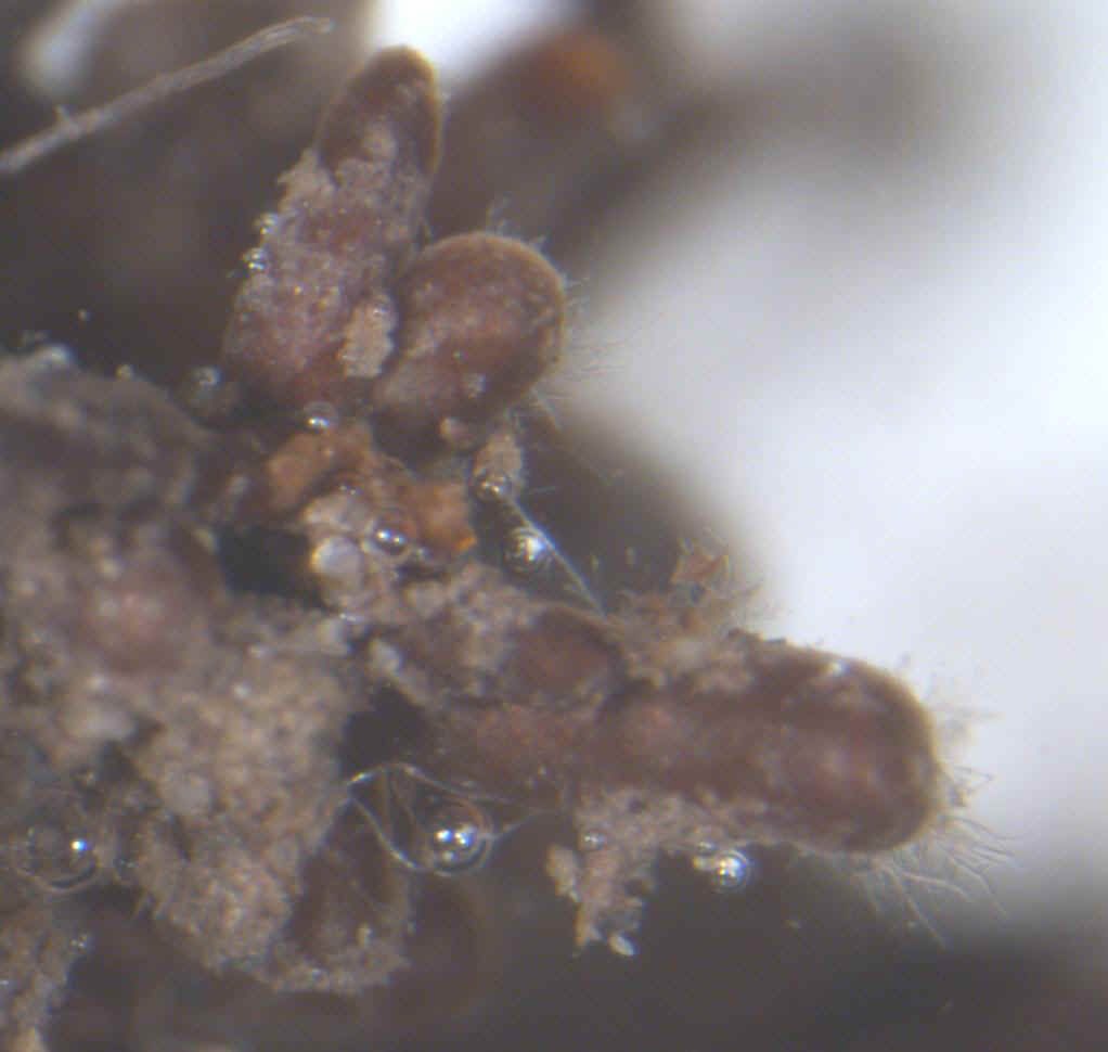
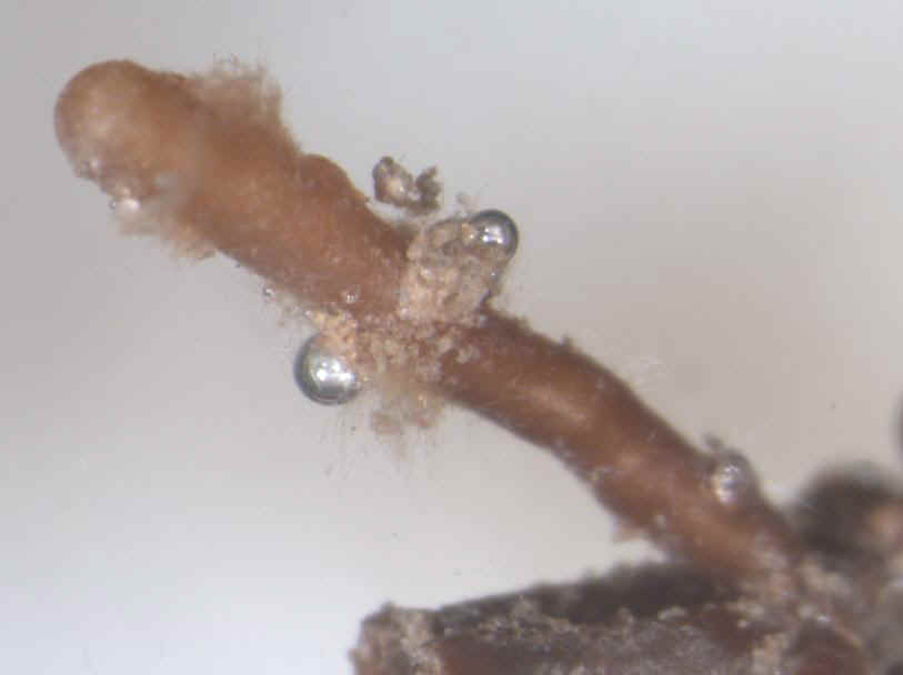
Figure 4. Dissecting microscope view (x25) of ectomycorrhizae characteristic
of Tuber melanosporum on Oak #4. Note the presence of characteristic emanating
hyphae (arrows), disqualifying the possibility of any other truffle species,
i.e. long, hyaline, loosely arranged, often branching at approximately right
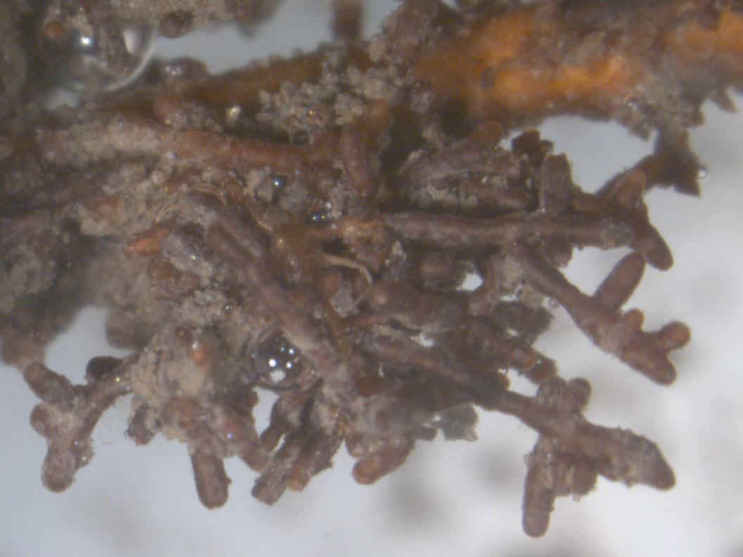
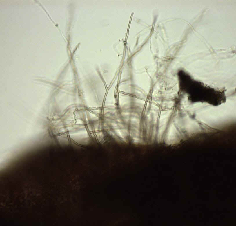
angles and never tapered at the apex (not ‘spiky’, see also Figure 6). Figure 5. Dissecting microscope view (x7) of a cluster of Tuber melanosporum ectomycorrhizae on Oak #4.
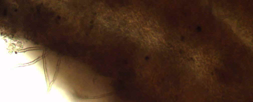
Figure 6. Compound microscope views (x300 left, x400 right) of emanating hyphae (Top) and of the ‘jigsaw puzzle’ pattern (Bottom right) of the mantle of ectomycorrhizae of T. melanosporum collected from Oak #4. The combination of these two characteristics is characteristic of T. melanosporum only. Emanating hyphae are long, hyaline, loosely arranged, often branching at approximately right angles and never tapered at the apex.
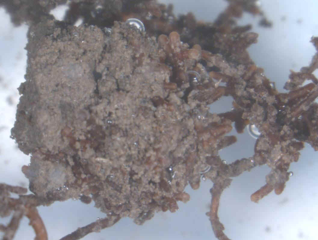
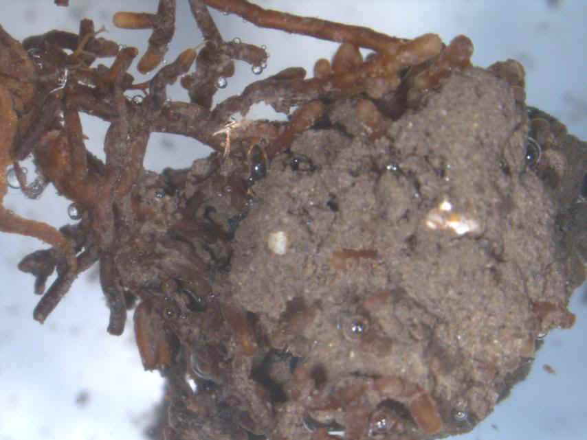
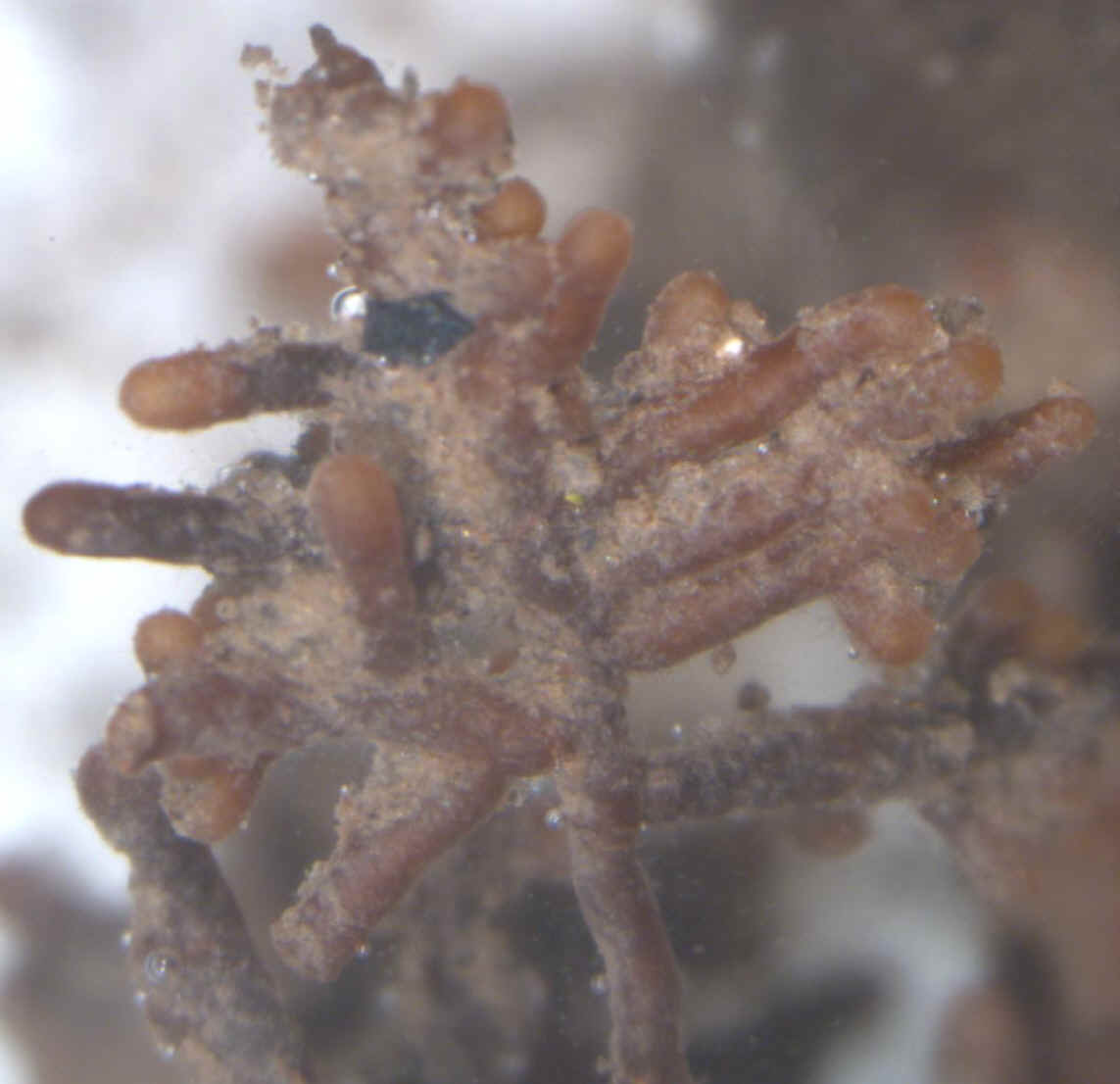
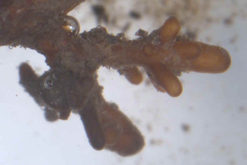
Figure 7. Dissecting microscope views of ectomycorrhizae (x15 top left, x7 bottom left and right) characteristic of Tuber melanosporum on Oak #5. Note the presence of well-developed clusters of multi-branched mycorrhizae (Bottom).
Figure 8. Compound microscope view (x400) of the jigsaw puzzle pattern of an ectomycorrhiza (left) characteristic of Tuber melanosporum on Hazel #6.
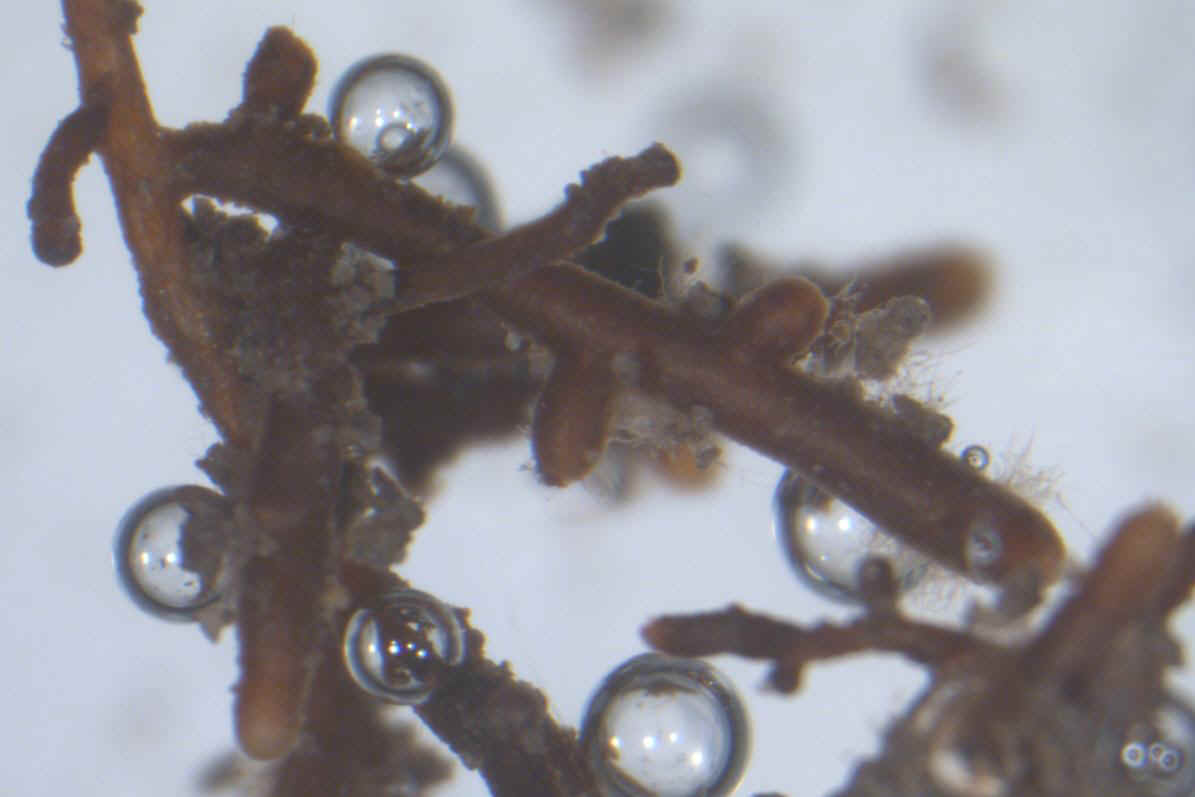
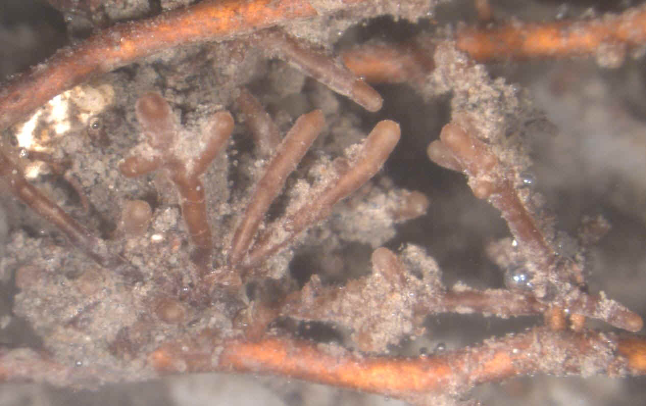
Figure 9. Dissecting microscope view (x12) of ectomycorrhizae (right) characteristic of Tuber melanosporum on Hazel #6. Note the presence of well-developed clusters of ectomycorrhizae.
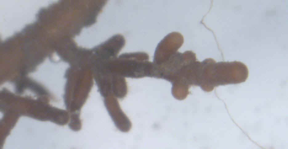
Figure 10. Dissecting microscope views (x10 left, x40 right) of ectomycorrhizae characteristic of Tuber melanosporum on Hazel #8. Note the presence of characteristic emanating hyphae (arrow).
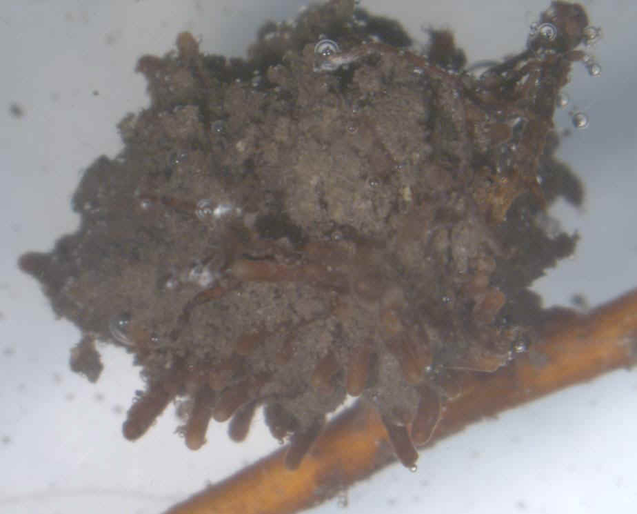
Figure 11. Unmagnified view of a very large cluster of ectomycorrhizae of Tuber melanosporum on Hazel #9 (Top left). Dissecting microscope view of a multi-branched ectomycorrhiza of T. melanosporum from Hazel #9 (x15 top right). Compound microscope view (x350) of both the jigsaw puzzle pattern (*) and characteristic emanating hyphae (arrow) from a T. melanosporum ectomycorrhiza of Hazel #9 (Bottom right). Both characteristics are unique to T. melanosporum and rare to capture on the same microscope slide.
Figure 12. Dissecting microscope views (x15 top left, x20 top right) of ectomycorrhizae characteristic of Tuber melanosporum on Hazel #10. Note the presence of well-developed clusters of ectomycorrhizae (x10 bottom right).
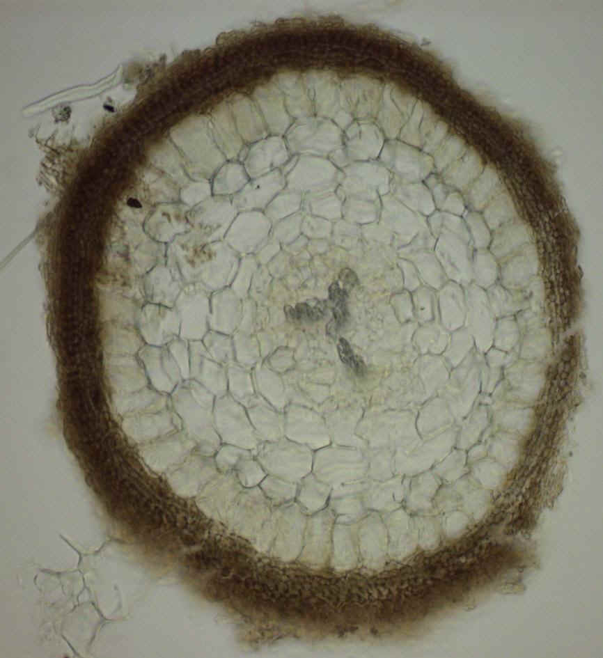
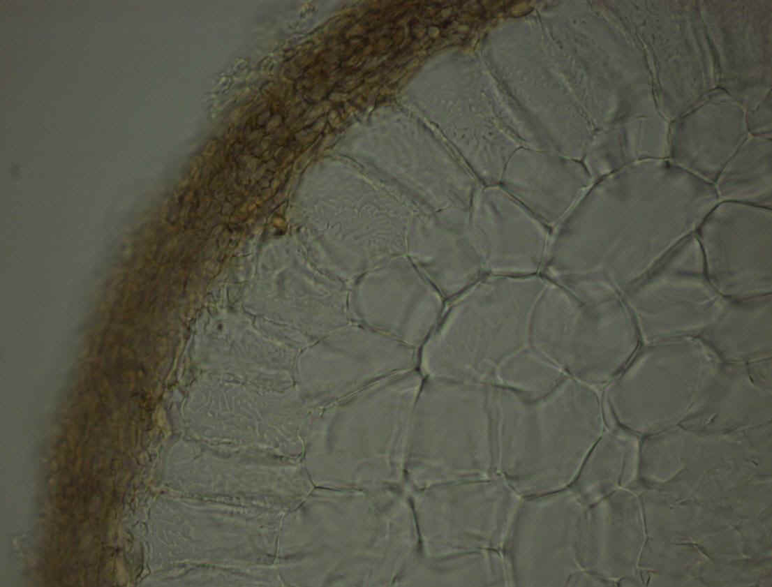
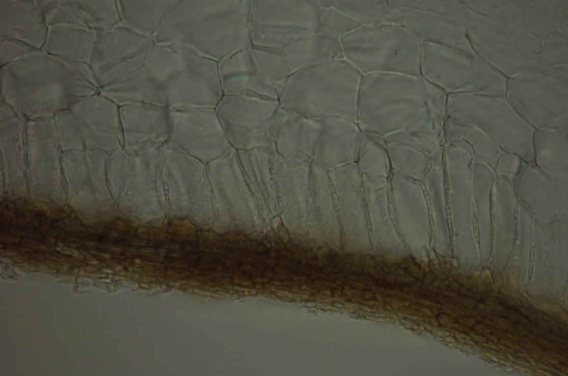
Figure 13. Compound microscope views (x150 top right, x400 right) of transversal (Top) and longitudinal (Bottom right) freezing microtome sections of un-stained ectomycorrhizae of Tuber melanosporum on Hazel #9. Note the thickness and multi-layers structure of the mantle. Note the well-developed labyrinthine ‘Hartig net’ of fungal hyphae (arrows) enveloping the root cortical cells (Top right). The one cell layer structure of the Hartig net is visible on the longitudinal section (arrow, Bottom right). Courtesy Wang Yun.
|
|